

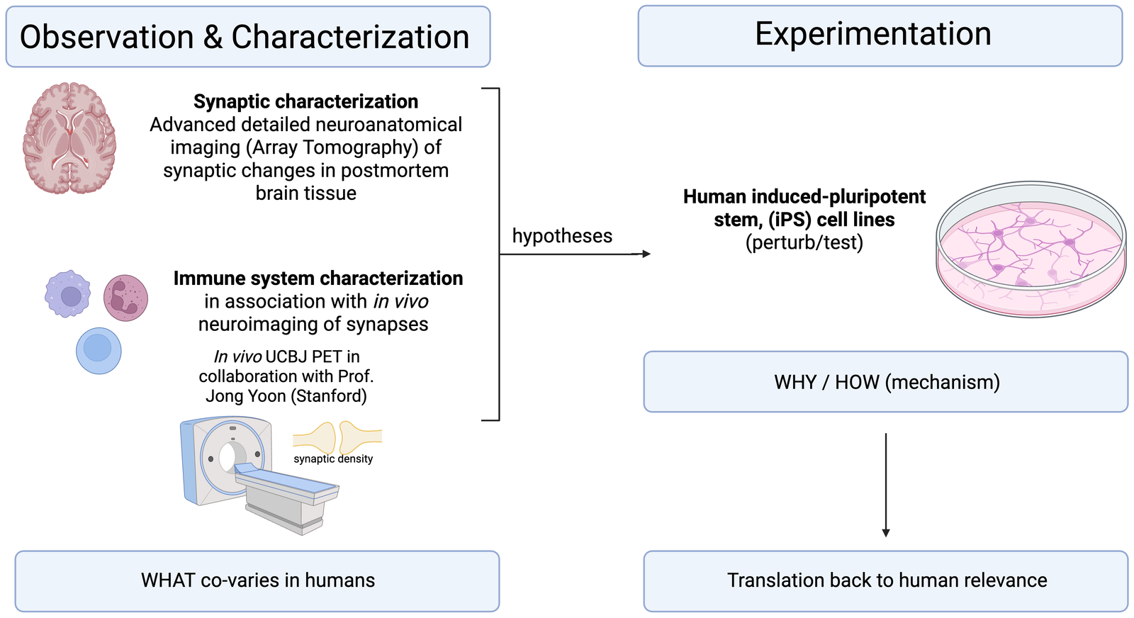
Converging evidence points to the synapse, the connection between neurons that forms the basis of cognition, as a primary site of pathology in schizophrenia. Our laboratory studies synaptic pathology through three complementary approaches: high-resolution imaging of postmortem brain tissue, mechanistic experiments in stem-cell models, and analysis of patient-derived blood to identify peripheral factors that shape synaptic pathology. Together, they move from characterizing what is altered in patients toward the mechanisms that cause it, with the goal of disease-modifying therapies.

Lab research overview: observation and characterization in human tissue and blood, and experimentation in iPS cell models.
Key discovery

Neutrophil C4 protein in schizophrenia

In brief: We identified neutrophils as a previously unrecognized potential source of complement activation in schizophrenia, and preliminarily linked neutrophil C4 protein to both clinical symptoms and in vivo synaptic density.

How it began

This work started in 2018 with a clinical observation: several patients with schizophrenia showed elevated complement component 4 (C4) activation fragments during extended immunological workups in which all other parameters were normal. This came shortly after the landmark 2016 Sekar et al. study, which showed that much of the schizophrenia genetic signal in the Major Histocompatibility Complex (MHC) could be explained by C4 gene copy number, and that C4 protein participates in synaptic pruning.

Confirming the signal

This observation led to a small funded observational clinical trial confirming that C4 activation products were elevated in individuals with schizophrenia [6]. Concurrently, other groups showed that complement activation in schizophrenia correlates with clinical symptoms, patient outcomes, and neuroimaging findings, yet does not follow traditional complement pathways.

Resolving the paradox

This inconsistency led to our central discovery: neutrophils, which make up roughly half of all white blood cells, express C4A mRNA and contain C4 protein [1]. We found that neutrophil C4 protein was:

  • Lower in schizophrenia samples than in controls, and correlated with C4A gene copy number, suggesting active expression and consumption with release of activation products into the blood [1]
  • Positively correlated with clinical measures in exploratory analyses, including a self-report stress measure and a clinician-rated symptom measure, supporting its link to disease status and its likely role as the source of complement activation
  • Negatively correlated with in vivo synaptic density in individuals with schizophrenia, in an overlapping cohort from our study [1] and Dr. Jong Yoon’s PET neuroimaging study of synaptic density [4] [manuscript in preparation]

Why it matters

This work links several previously disconnected aspects of schizophrenia pathophysiology:

  1. C4 gene copy number confers disease risk
  2. Neutrophils are both elevated and activated in patients
  3. Clozapine, the most efficacious treatment for schizophrenia, inhibits neutrophils
  4. Neutrophil C4 protein is the likely source of complement activation
  5. Neutrophil C4 protein tracks with clinical measures and neuroimaging-based synaptic density

Together, these findings reveal a source of complement activation occurring outside the canonical plasma-based complement cascade, and suggest that neutrophil C4 protein may directly modify synapses in schizophrenia, or reflect synaptic status.

Ongoing & future work

Where the lab is headed

Characterizing immune mechanisms driving synaptic alterations

Central question. Is neutrophil C4 protein a direct contributor to disease (a therapeutic target) or a consequence of it (a biomarker)?

Illustration of proteins at a synapse between neurons, depicting molecular signaling at the neural connection.

To test whether neutrophil C4 drives synaptic pathology, we pursue four complementary lines of work.

Clinical validation

Confirm the neutrophil C4–synaptic density correlation in a larger cohort.

Neutrophil biology

Detail the role of C4 protein in neutrophils using a neutrophil cell line and isolated primary neutrophils.

Donor profiling

Perform deep proteomic and transcriptomic profiling of neutrophils from schizophrenia and control donors.

Synaptic mechanism

Determine the molecular signals required for neutrophils to modify synapses, using a genetically engineered iPSC-based in vitro model.

These experiments address long-standing field questions: whether peripheral immune activation detrimentally impacts the brain, and whether inhibiting neutrophil activation confers therapeutic benefit.

A second immune mechanism: C4A upregulation

The cytokines IFN-γ and IL-6, both elevated in the schizophrenia periphery and cerebrospinal fluid, upregulate C4A expression. While elevated C4A is thought to drive excessive synaptic pruning, a large multisite trial blocking microglial activation failed to show benefit, and postmortem studies find no evidence of microglial activation [7,8], pointing to a mechanism beyond microglia.

In an upcoming manuscript, we will show how C4A protein has functions beyond synaptic pruning. [manuscript in preparation]

From patient-derived evidence to mechanistic models

Central question. Is neutrophil C4 protein a direct contributor to disease (a therapeutic target) or a consequence of it (a biomarker)?

Illustration of proteins at a synapse between neurons, depicting molecular signaling at the neural connection.

To test whether neutrophil C4 is a driver of synaptic pathology, our work moves from patient-derived evidence toward mechanistic models.

Human evidence

We are confirming the neutrophil C4–synaptic density correlation in a larger cohort and performing deep proteomic and transcriptomic profiling of neutrophils from schizophrenia and control donors.

Mechanistic models

We are detailing the role of C4 protein in neutrophils using a neutrophil cell line and isolated primary neutrophils, and determining the molecular signals required for neutrophils to modify synapses in a genetically engineered iPSC-based in vitro model.

These experiments address long-standing field questions: whether peripheral immune activation detrimentally impacts the brain, and whether inhibiting neutrophil activation confers therapeutic benefit.

Detailing synaptic architecture using Array Tomography

A core platform of the lab uses Array Tomography, an advanced multiplex spatial imaging technique developed at Stanford, to characterize synapses in postmortem brain from individuals with schizophrenia versus controls [NIH K08, funded through 2/2029].

Diagram of the Array Tomography workflow: serial ultrathin brain sections, multiplex fluorescent imaging, and 3D reconstruction for synaptic analysis.

Testing the excitatory–inhibitory imbalance hypothesis

The current project establishes this technique in a large biobank cohort by measuring inhibitory and excitatory synaptic density in affected regions. Because simultaneous measurement of both major synaptic subtypes has never been achieved due to technical limitations, doing so will directly test the excitatory–inhibitory imbalance hypothesis in schizophrenia.

Building a resource for the field

More complex synaptic characterization will follow in regions of interest, creating a detailed resource for understanding synaptic composition in schizophrenia, extensible to other psychiatric and neurodegenerative disorders.

Connecting structure to mechanism

Array Tomography will be coupled to other datasets to validate mechanisms and their association with synaptic pathology in clinical brain tissue.